Deep Pulp Chamber
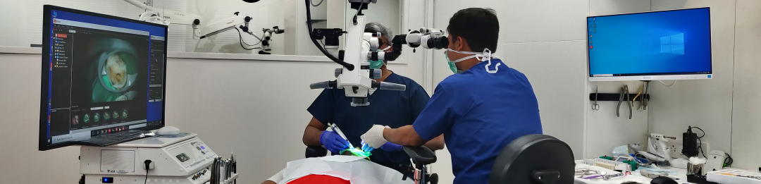
This was a case where there was a large cervical caries leading to pulpitis. The pulp chamber was calcified and the floor was quite deep. Fortunately, the microscope makes these cases a bit more predictable.
This was a case where there was a large cervical caries leading to pulpitis. The pulp chamber was calcified and the floor was quite deep. Fortunately, the microscope makes these cases a bit more predictable.
This is a garden variety middle-mesial canal i tretaed in 2009. The crown done by the referral was a bummer. Patient came back a year later with discomfort. The periapical lesion had healed, but the crown had an open margin leading to food impaction. I suggested changing the crown. Liviu Steier requested that I convert […]
This is a case from 12 years ago. I re-treated all the canals including the distal which had a metal post. At, the one-year recall, the mesial root seems to have healed well. Today, I would probably have left the distal root alone and not over-enlarged those mesial canals.
This is a case from the old archives. I used to get a lot of these before 2010. I don’t see many silver cones anymore. I used to use this as a teaching case for how to remove silver cones.
This was an old case i discovered on my comp. I treated this case in 2006. It is probably a good case to demonstrate the location of the middle mesial canal.Strangely, the first thing that I thought when I saw this case today was, “Did this really benefit the patient? There wasn’t any lesion and […]
This was one of the first apexification cases i did. I started this case before i purchased a microscope (and I wasn’t using MTA at that time). I packed the canal with a thick Iodoform + Calcium hydroxide (Metapex). By the time the patient came back after a year, I had a microscope and it […]
Here is a step by step pictorial guide to locating a calcified canal in a mandibular premolar. Pictures show colour differentiation between calcifications and dentine.
This was a routine Maxillary pre-molar Re-treat. Always nice to see some anatomy being filled in the end.
We often see maxillary second molars, which, at first glance seem to have just 2 canals, one Buccal and one palatal. A closer examination of the Buccal canal will often reveal bifurcation into two canals, DB and MB. Here is one such case.
This patient came with an acute abscess. Disassembly and re-treat with long term Caoh. One year recall shows healing.
