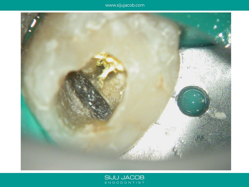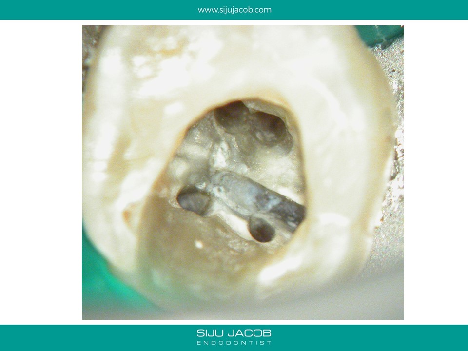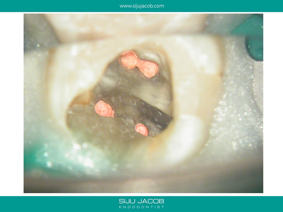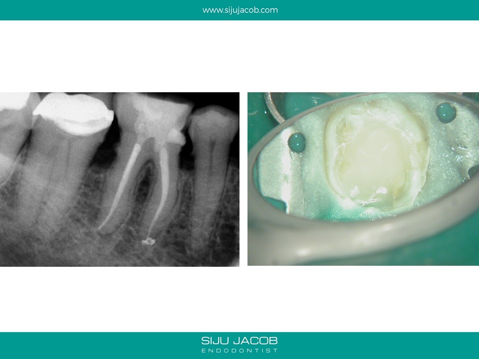
This case illustrates two things.
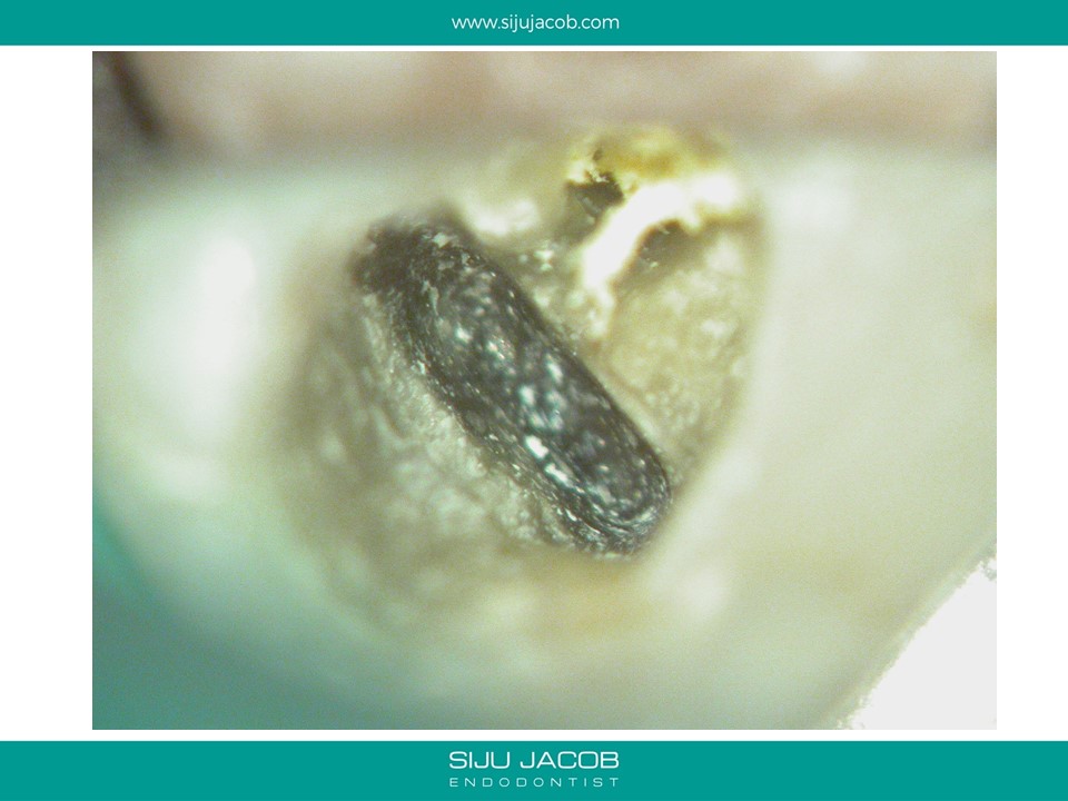
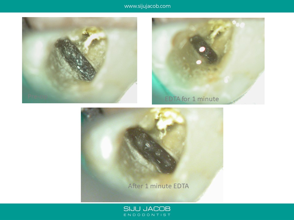
1) When examining for calcified canals, I like to put some EDTA, scrub the chamber and then examine under the microscope for clues. A cleaner floor is easier for color differentiation.
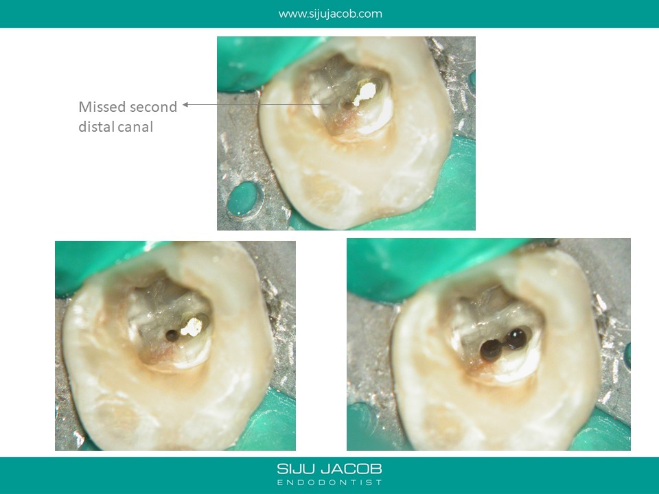
2) Specially in posterior teeth, canals tend to calcify and appear as “white spots”